Lameness
Lameness can be a complex puzzle and may involve the need to use special diagnostic and imaging techniques.
It is for that reason that many clients and / or referring veterinarians, send their difficult cases in to our hospital for further diagnosis and treatment. Here at GVEH we specialise in the diagnosis and management of lame horses- be that a racehorse, performance horse or paddock pony.
We can use a range of diagnostics including flexion tests, nerve blocks, radiographs, ultrasound and scintigraphy to get to the bottom of the problem.
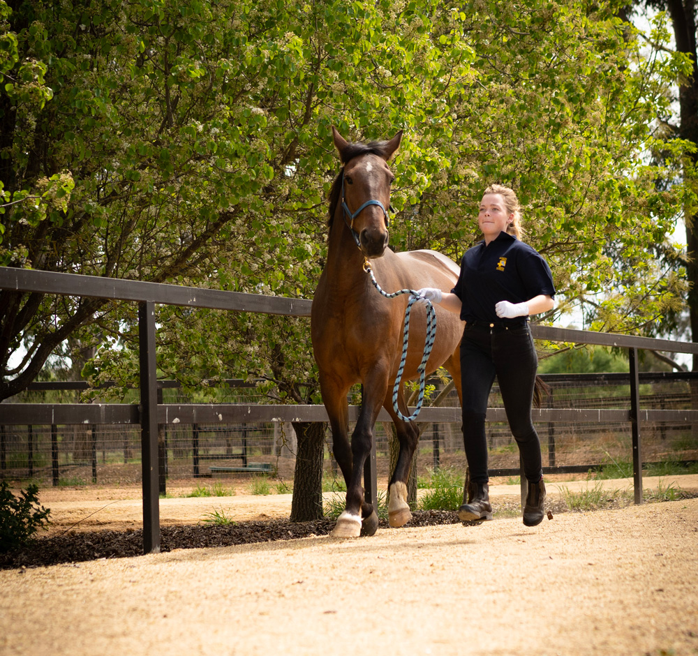
We have a specific lameness area with a hard, smooth surface that horses can be trotted on and two round yards (one with a hard surface and one with a soft).
These specifically designed areas aid our team to correctly assess your horse’s lameness at various gaits.
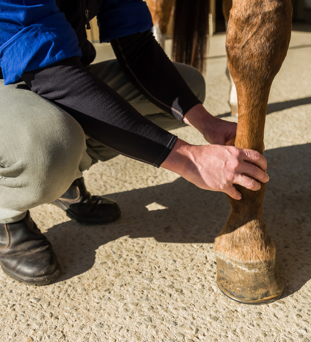
The flexion test involves holding the horse’s leg in a prolonged flexion then trot the horse away. The purpose is to accentuate any pain that may be associated with a horse’s joint or soft tissue structures. We can separate the horse’s limbs into different areas (lower, upper or full limb) this gives guidance of where to start with nerve or joint blocks.
To perform a nerve block, a small amount of local anaesthetic is used to anaesthetise (numb) nerve’s that provide sensation to various areas of the horse’s leg.
This is a short-term block and allows the team to assess which area the horse the lameness is and if any other areas of the horse maybe contributing to the current lameness.
At GVEH we use a combination of nerve and joint blocks to correctly identify the primary cause of lameness.
After the origin of lameness has been identified, X-rays, ultrasonography or other imaging techniques are used to best determine the diagnosis. From here we can guide you with the best way to treat your horse’s lameness.
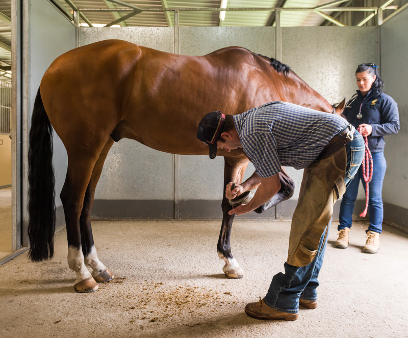
No dedicated lameness facility would be complete without involvement of an expert farrier. Our farrier is integral to our team approach.
We have a forge and room devoted to the manufacture of shoes and shoeing of horses with problems such as founder (laminitis), navicular disease, hoof lacerations as well as the manufacture of treatment plates and specialty shoes for surgery patients.
Our goal is to return your horse to their intended use as soon as it is safely possible.
Our holistic, team approach helps to achieve this goal.
In order to give us the best chance of seeing the cause of your horses lameness, we have the following tools at our disposal:
Radiography
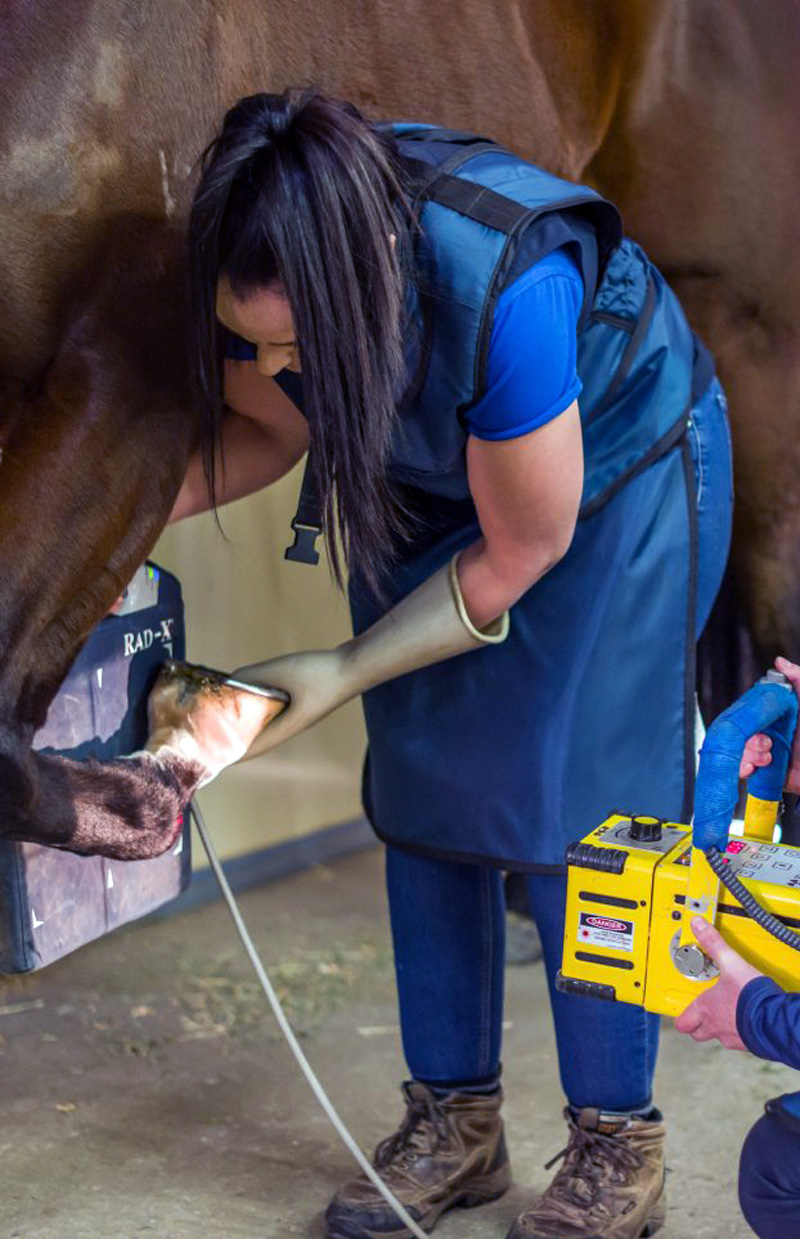
Has quickly become one of the most commonly used imaging modalities in our hospital. At GVEH we have two portable X-ray units which can be used around the hospital or taken on farm. Images are then sent through to a larger high-resolution screen in the hospital to enable our veterinarians to closely examine any abnormalities. Radiographs can show a range of issues ranging from joint disease, bone chips and fragments, osteochondritis disease (OCD), sequestrum and fractures, as well as some soft tissue abnormalities.
X-rays are generally taken based either on abnormal clinical findings, such as a swollen or painful joint, or after localisation using nerve blocks.
Because everything in the path of an x-ray beam ends up on the final picture, there is a great degree of overlay of structures. For this reason, several pictures of a joint are taken at different angles, so that different areas may be highlighted. Because of radiation hazard, and cost considerations, speculative x-raying of an entire limb is impractical, and generally unrewarding. This is especially true in older horses, which often have radiographical signs of wear and tear, which may not be associated with lameness.
One of the biggest limitations of radiology in horses is that x-rays cannot penetrate through large distances of muscle or bone, and so, with some selected exceptions, radiography of the abdomen and pelvic area is usually unrewarding in adult horses. When your horse has x-rays taken, we will often sedate him/her, as movement blur makes x-rays unreadable, and personnel are often placed in positions of potential danger in order to get diagnostic views.
Scintigraphy
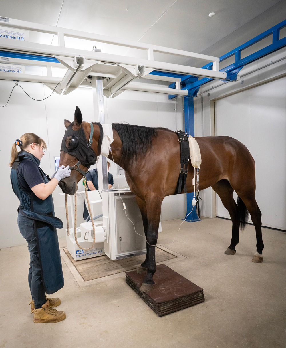
Is a highly sensitive imaging technique that allows our qualified staff to detect lameness injuries that have been difficult to diagnose by creating bone scans with our new state-of-the-art MIE Equine Scanner H.R. Scintron Gamma Camera.
After the horse is injected with radioactive dye that binds to injured bone, a large gamma camera produces images that highlight injury hot spots. These images are invaluable to making a correct diagnosis and developing an effective treatment plan.
Nuclear scintigraphy is particularly useful for diagnosing stress fractures where repeated jogging of the horse would be contraindicated, cases where diagnostic analgesia diagnosis (nerve and joint blocks) has been unsuccessful, or for horses that are dangerous to block.
The radioactive dye is completely safe and works out of the horse’s system within 24 hours.
Ultrasonography
Or ultrasound as it is commonly referred to, is the use of sound waves with frequencies higher than the upper audible limit. It is a valuable diagnostic tool used commonly to image soft tissue structures such as tendons and ligaments. Ultrasound can also be used to aid the examination of the reproductive tract, all joints and bone surfaces. Ultrasound can be helpful for assessing any upper limb injuries such as pelvis or shoulder injuries that cannot be x-rayed.
Specialist Farrier
No dedicated lameness facility would be complete without involvement of expert farrier attention. Our farrier is integral to our team approach. We have a forge and room devoted to the manufacture of shoes and shoeing of horses with problems such as founder (laminitis) or navicular disease.
One of the goals of our veterinary attention is to return the horse to work as soon as safely possible after the problem has been corrected. Attention to proper therapeutic shoeing reduces this time.
We have a specialist farrier on site every Tuesday who takes regular bookings for corrective trim/shoeing.
